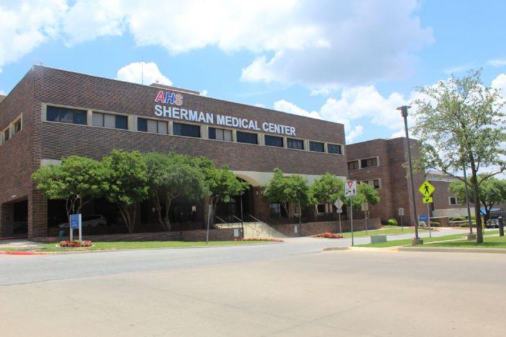
Next week, American Healthcare Systems Sherman Medical Center will be opening a new department. The former Wilson N. Jones Hospital, will open its new women’s imaging center the first week of November but will celebrate the new office with a ribbon cutting to be held Wednesday.
Scheduling has already started with more than 100 appointments on the books.
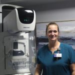
“Scheduling is very busy right now,” Center Manager Rebekah Clements said Wednesday. “I think we had 130 initial employees and family members who we let sign up first. They are our first patients and then from there we will be completely open to the public. We have quite a few scheduled and we are excited.”
Clements has been a mammographer for 17 years. She is also a certified breast ultrasound tech. She will be joined in the office by a volunteer who will great patients along with a registration person and mammographer Tricia Phillips who has been working in the field for 15 years.
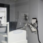
“This area used to be a GI lab,” Clements said as she gave a tour of the new office located just inside AHS Sherman Medical on the first floor. “This is a very big area from where I have worked breast center-wise. The rooms in this facility are quite large.”
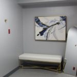
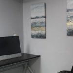
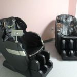
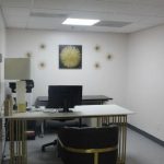
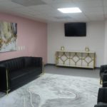
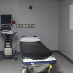
With couches, a refrigerator and massage chairs awaiting the first patients, Clements said hospital leadership wanted to make sure the new center was welcoming for those seeking services.
“We do not want this area to feel sterile and overwhelming because this is tough enough in itself,” she said. “People get so nervous over mammography and I understand that. If you have a history of it or this is your first time or you have had an abnormal mammogram then it does feel overwhelming. So, we wanted the environment to feel welcoming. We want it to be a safe place, a quiet place, a welcoming place.”
The center will offer screening mammograms, diagnostic imaging and ultrasounds.
“There will be screening mammograms everyday, and then we will have diagnostic imaging which means any patient with a problem, or with a history of breast cancer in the last five years, they will be imaged diagnostically on Thursdays,” Clements said. “We will have a radiologist that is available to us on those days that we will be able to connect with to get a result right then.”
That will be from about 8 a.m. to noon every Thursday.
“We will eventually have the ability to biopsy here,” Clements said. “We are still working to that.”
For anyone wishing to have a screening mammogram, the books are open now and a referral from a doctor is not needed.
“Screening mammograms do not require a doctor’s order,” Clements said. “As long as your are not having a problem, it does not require a referral. But if you are having discomfort, discharge, thickening or redness and warmth, those require a diagnostic mammogram which is a doctor’s order.”
This will not be your grandmother’s mammogram.
“Mammography is not what it used to be,” Clements said. “It is still intense and cold. The plate is always going to be cold. But the mammogram is much more comfortable than it used to be. We even have the curved paddles that are even better. The compression is better. It does not require as much. The imaging is better. You can breathe through the pictures now. It can be less overwhelming. This process is not near where it used to be.”
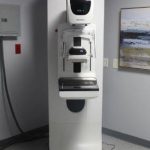
The hospital got some new equipment to make sure the services will be as up to date as possible. The Selenia 3D imaging system is one of the latest machines used for breast imaging technology.
“So, it takes images at slices throughout the breast tissue,” Clements said. “So, if you can compare it to like a CAT scan. The radiologist is able to scroll through those images and see layer by layer.
“So, it has been described to me like with 2D imaging it is like looking at the front and back cover of a book. This kind of 3D imaging is like being able to see the front and back cover along with all the pages in between. They can see every layer of your breast tissue and be able to catch the smallest things.”
This can help also with imaging in the past that seemed to be more difficult.
“This can be particularly important for dense breast tissue, but we have seen it work wonders on in fatty breast tissue too,” she said. “It is phenomenal technology. It really blows me away. I get geeked out over it because of what they can see now. I tell people all the time all day that this is not how it used to be when it comes to having a mammogram. This is not your grandma’s mammogram. This is newer. It is advanced technology and not as uncomfortable as it used to be.”
And that does not just apply to women. Anyone can be screened for breast cancer.
“Be aware that screening is our best line of defense against breast cancer,” she said. “We cannot stop you from getting breast cancer, but if you screen regularly, we can catch it as early as possible. That is the best prevention. That is best type of detection, early detection.”
And routine screenings are important because they help doctors see what changes are taking place over time.
“This is also a way to build a portfolio because the doctor can look back at your past screenings and see what was normal and the changes to know what is abnormal for you,” she said. “That way they can catch it as early as possible.”
Clements also said that a misconception is that a patient should get an ultrasound over a mammogram.
“Breast ultrasound is to be used in conjunction with mammography,” she said. “It is something to be utilized with mammography. It is not stand alone. Sometimes people come in saying they want an ultrasound and not a mammogram. You do not want to do that. The earliest signs of breast cancer are calcification. We do not see those on ultrasounds. Ultrasounds are not the best tool. Mammograms are, but ultrasounds with mammography provide a great picture.
Individuals can start getting mammograms at 35 depending on person history or family history. For more information or to reach the hospital, call 903-870-4611.